 +971 6 522 6368
  +971-56-994-6523
  info@resalahphysio.com
Acromioclavicular (AC) Joint Injury Physiotherapy & Rehabilitation
1. Introduction & Anatomical Overview
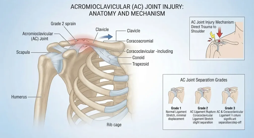
The Acromioclavicular (AC) joint is a common site of injury, particularly for athletes involved in contact and collision sports or activities that involve frequent throwing. Anatomically, the AC joint is a crucial component of the shoulder structure, located at the point where the lateral end of the clavicle (collar bone) meets the acromion process of the scapula (shoulder blade). It can be easily identified by sight and touch as the pointy protrusion near the top, outer edge of the shoulder.
The joint is entirely enclosed by a joint capsule and receives primary structural support from the acromioclavicular and coracoclavicular ligaments. In most typical AC joint injuries, these specific stabilizing ligaments undergo micro-tearing, partial disruption, or complete rupture.
At Al Resalah Medical Center, our multidisciplinary team approaches AC joint injuries with a rigorous clinical eye, understanding that accurate classification and structured rehabilitation are vital to restoring full upper extremity biomechanics and preventing chronic shoulder instability, persistent pain, or post-traumatic degenerative changes.
2. Classification & Grading of Ankle Sprains
Grade 1 (Mild)
Pathology: Partial damage/microscopic stretching to the joint capsule and AC ligament; no macroscopic tear of the coracoclavicular ligament.
Clinical Presentation: Mild localized tenderness, discomfort or pain when the clinician assesses and moves the joint. Full functional range of motion is largely preserved.
Expected Recovery: 1 to 3 weeks.
Grade 2 (Moderate)
Pathology: Complete rupture of the acromioclavicular ligaments combined with partial tearing of the coracoclavicular ligament.
Clinical Presentation: Moderate to severe pain, localized swelling, restricted shoulder movement, and an upward displacement of the clavicle causing a noticeable step-deformity or larger bump.
3 to 6 weeks
Grade 3 (Severe)
Pathology: Complete rupture of both the acromioclavicular and coracoclavicular ligaments, resulting in complete joint capsule dislocation.
Clinical Presentation: Severe immediate pain, extensive soft tissue swelling, profound mechanical instability, and an obvious, prominent lump at the end of the collar bone.
Expected Recovery: 6 to 12+ weeks (Surgical consultation may be indicated depending on patient functional demands).
3. Symptoms and Etiology
Understanding the precise mechanism of injury provides critical diagnostic insight. AC joint trauma generally occurs via two primary pathways:
Direct Trauma: A direct blow to the tip of the shoulder (e.g., an awkward fall onto the lateral shoulder or impact with another player), which forces the acromion process downward beneath the clavicle.
Indirect Trauma: An upward force transmitted along the long axis of the humerus, such as a fall directly onto an outstretched, straightened arm while the shoulder is positioned close to the body (adducted) and flexed.
Common Symptoms
Acute, localized pain directly at the distal end of the collar bone.
Widespread shoulder discomfort initially, which gradually localizes specifically to the AC joint as acute inflammation resolves.
Localized soft tissue swelling and ecchymosis around the joint line.
Visible step-deformity or pronounced lump at the outer edge of the shoulder (depending on injury grade).
Restricted and painful shoulder movement, especially when attempting to elevate the arm at or above shoulder height.
Primary Etiological Factors
Participation in contact, collision, or overhead throwing sports.
History of prior shoulder girdle trauma or previous AC joint sprains.
Deficits in dynamic scapular stability and rotator cuff strength.
Lack of appropriate protective padding or strapping during high-risk athletic competition.
4. Clinical Diagnosis & Diagnostic Imaging
A comprehensive physical examination performed by our experienced medical practitioners and neuro-physiotherapists at Al Resalah Medical Center forms the bedrock of accurate diagnosis. Physical assessment includes evaluating active and passive range of motion, neurovascular status, and specific orthopedic provocation tests (such as cross-body adduction tests and palpation of the AC joint line) to isolate pathology from glenohumeral or cervical spine conditions.
Diagnostic Imaging & Clinical Efficiency
To ensure accurate grading and rule out associated clavicular or scapular fractures, standard radiographic evaluations (such as weighted or unweighted anteroposterior and Zanca views of the clavicles) are utilized when clinical presentation warrants imaging. Our clinicians adhere to strict evidence-based guidelines to ensure appropriate diagnostic screening.
5. Management and Treatment Framework
Modern clinical management of AC joint injuries emphasizes acute protection followed by progressive, active rehabilitation. At Al Resalah Medical Center, our treatment protocol is structured into three distinct phases designed to restore full functional independence.
Phase 1: Acute Protection & Pain Modulation (Days 1 to 7)
RICER Protocol: Rest, Ice, Compression, Elevation, and Referral. The shoulder should be rested in an elevated position with an ice pack applied for 20 minutes every 2 hours (avoiding direct skin contact) for the first 48 to 72 hours to control hemorrhage, reduce swelling, and minimize tissue damage.
Immobilization: The arm is rested in a supportive sling during the acute window to immobilize and prevent harmful micro-movement.
No Harm Protocol: Strict avoidance of heat, alcohol, physical running/activity, and manual massage to the acute site.
Modalities & Gentle Mobility: Targeted use of non-invasive electrophysical agents and early, pain-free passive range of motion as tolerated.
Phase 2: Restoration of Range of Motion & Neuromuscular Control (Weeks 2 to 4)
Manual Therapy: Gentle joint mobilizations and soft tissue techniques to address secondary cervical and shoulder girdle stiffness without compromising ligament healing.
Progressive Strengthening: Initiation of sub-maximal isometrics progressing to light resistance training for the rotator cuff, deltoid, and scapular stabilizers.
Scapular Kinematics: Retraining proper scapulothoracic movement patterns to ensure harmonious joint mechanics during arm elevation.
Phase 3: Functional Retraining & Sport-Specific Conditioning (Weeks 5+)
Advanced Strengthening: Closed and open kinetic chain exercises designed to rebuild structural resilience across the shoulder complex.
Sport-Specific Simulation: Controlled functional drills, gradual re-introduction of throwing or contact mechanics, and application of protective athletic strapping where necessary.
6. Al Resalah Medical Center Physiotherapy Specialists
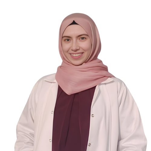
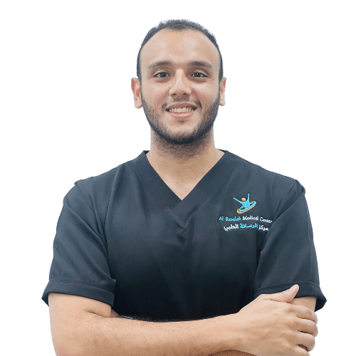
Our dedicated rehabilitation department features highly qualified clinicians specializing in musculoskeletal, neurological, and sports physiotherapy. Every patient receives an individualized, evidence-based care plan supervised by our senior specialists:

7. Advanced Physiotherapy Treatments
To accelerate cellular healing, reduce recovery times, and ensure superior clinical outcomes, Al Resalah Medical Center utilizes cutting-edge therapeutic technologies:
Extracorporeal Shockwave Therapy (ESWT): High-energy acoustic waves directed into damaged ligamentous tissue to stimulate local healing responses and decrease chronic inflammatory markers.
High-Intensity Laser Therapy (HILT): Penetrates deep into soft tissues to stimulate mitochondrial cellular activity, accelerate tissue repair, and provide rapid analgesic relief.
Instrument-Assisted Soft Tissue Mobilization (IASTM): Specialized instruments used to manage fascial restrictions and fibrotic tissue changes following shoulder trauma.
Clinical Warning: Avoid Premature Return to Activity A common pitfall in AC joint recovery is returning to heavy lifting, contact sports, or overhead activities prematurely simply because superficial surface pain has subsided. Incomplete ligamentous healing or unaddressed scapular dyskinesis drastically increases the risk of chronic joint instability and persistent discomfort. Always complete the full functional progression under professional guidance.
8. Common Home Exercise Program (HEP)
To support in-clinic rehabilitation, patients are prescribed a customized home exercise program. Consistency is vital for achieving full functional recovery.
1. Pendulum Exercises (Early Mobility)
- Execution: Lean forward slightly, supporting your uninjured arm on a table. Let your injured arm hang completely relaxed downward. Using your body momentum, gently swing the arm in small circles, side-to-side, and front-to-back.
- Sets/Reps: 2–3 sets of 1–2 minutes, 2 to 3 times daily.
2. Isometric Shoulder Internal/External Rotation
Execution: Stand near a doorframe or wall with your elbow bent at a 90-degree angle. Place your hand against the wall attempting to push outward (external rotation) or inward (internal rotation) without actually moving your arm. Hold a sub-maximal contraction.
Sets/Reps: 3 sets of 10 holds (holding each for 5 seconds).
3. Scapular Squeezes (Postural Control)
Execution: Sit or stand tall with arms relaxed by your sides. Squeeze your shoulder blades together and downward, holding for a few seconds before relaxing.
Sets/Reps: 3 sets of 10–12 repetitions, twice daily.
4. Resistance Band Rows (Scapular Stabilization)
Execution: Secure a resistance band at chest height. Hold the handles with elbows bent at your sides, and pull the band backward by squeezing your shoulder blades together, keeping your posture upright. Return with control.
Sets/Reps: 3 sets of 10–12 repetitions.
Written by Fayyaz Husain Physiotherapist
Medically Reviewed by Al Resalah Medical Team
Last Reviewed: July 2026