 +971 6 522 6368
  +971-56-994-6523
  info@resalahphysio.com

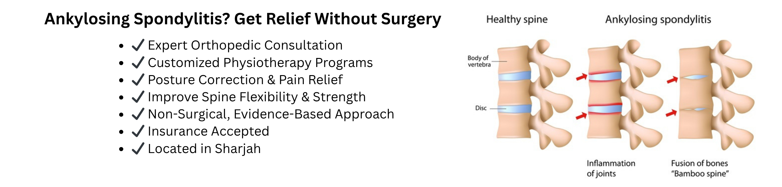
Ankylosing Spondylitis (Non-Surgical Management) Treatment in Sharjah
1. Introduction & Anatomical Pathology
Ankylosing Spondylitis (AS) represents a chronic, progressive, and systemic autoimmune-mediated inflammatory arthropathy targeting the axial skeleton. At Al Resalah Medical Center in Sharjah, our multidisciplinary team approaches this complex condition through a rigorous neuro-musculoskeletal and orthopedic lens. We recognize that inadequate management is a primary driver of irreversible spinal structural deformity, severe functional disability, and diminished pulmonary capacity.
Anatomically, AS selectively attacks the entheses—the specialized functional junctional zones where ligaments, tendons, and joint capsules insert directly into the bone matrix. Consequently, the primary sites of initial pathological involvement include:
Sacroiliac (SI) Joints: The crucial biomechanical link between the spine and pelvis. These joints almost universally exhibit the earliest inflammatory changes, known as bilateral sacroiliitis.
Zygapophyseal (Facet) Joints: The true synovial joints between adjacent vertebral arches. These structures dictate spinal rotation and gliding mechanics.
Costovertebral and Costotransverse Joints: The critical skeletal articulations connecting the thoracic vertebrae to the rib cage. These are vital for thoracic excursion and diaphragmatic breathing mechanics.
Annulus Fibrosus of Intervertebral Discs: Sites where chronic inflammation instigates fibrovascular tissue proliferation. Ultimately, this leads to pathological endochondral ossification. Over time, this biological repair mechanism results in bridging syndesmophytes—bony bridges fusing adjacent vertebrae into a rigid, unified column classically described as a “bamboo spine.”
2. Pathophysiological Staging & Disease Progression
To establish targeted non-surgical intervention strategies, our clinicians categorize Ankylosing Spondylitis progression through clear pathophysiological phases:
Stage 1: Active Inflammatory Phase (Synovitis & Enthesitis)
Cellular Mechanism: Pro-inflammatory cytokines, including Tumor Necrosis Factor-alpha and Interleukin-17, infiltrate synovial and entheseal tissues. This action provokes local edema, vascular congestion, and intense localized pain.
Clinical Presentation: Patients report severe morning stiffness exceeding 45 minutes, a deep-seated gluteal or lumbar ache, and systemic fatigue without definitive radiographic bone destruction yet visible.
Rehabilitation Focus: Clinicians emphasize active inflammatory suppression, pain modulation, preservation of joint range of motion, and avoidance of sustained spinal flexion positions.
Stage 2: Fibro-Osseous Proliferation & Structural Remodeling
Cellular Mechanism: Granulation tissue replaces eroded bone and cartilage at insertion sites. Afterwards, abnormal osteoblast activation occurs alongside calcification of the outer intervertebral disc fibers.
Clinical Presentation: Individuals experience a loss of normal lumbar lordosis, flattening of the thoracic curve, measurable reductions in spinal flexion and extension, and localized mechanical stiffness.
Rehabilitation Focus: Programs prioritize intensive axial extension therapy, posture re-education, maintenance of thoracic wall compliance, and anti-osteoporotic preventative conditioning.
Stage 3: Complete Ankylosis & Structural Fusion
Cellular Mechanism: Mature trabecular bone bridges across joint spaces, entirely eliminating micro-motion. Consequently, vertebrae stabilize into a rigid structural unit.
Clinical Presentation: Patients suffer a total loss of spinal mobility in affected segments, a pronounced forward-stooped hyper-kyphotic posture affecting horizontal gaze, and severe restriction of chest wall expansion during forced inspiration.
Rehabilitation Focus: Protocols center on fall prevention strategies, adaptation training for activities of daily living, and advanced respiratory physiotherapy.
3. Symptoms, Clinical Presentation & Differential Etiology
Understanding the precise clinical profile of inflammatory back pain versus mechanical spinal pathology remains critical for accurate diagnosis.
Primary Clinical Manifestations
Inflammatory Spinal Pain: Patients experience deep-seated, insidious lower back and buttock pain manifesting before age 45 and persisting beyond three months.
Paradoxical Response to Activity: Unlike mechanical disc or facet pain—which worsens with physical exertion and improves with rest—AS pain and stiffness worsen significantly with prolonged rest or immobility. Remarkably, they ameliorate with physical exercise.
Nocturnal Pain Wakening: Severe middle-of-the-night or early morning discomfort frequently forces patients to arise and walk around to ease symptoms.
Peripheral Enthesitis and Arthritis: Up to 30% of cases feature asymmetric involvement of peripheral joints like the hips, knees, and shoulders, alongside localized tenderness at the Achilles tendon insertion or plantar fascia origin.
Extraskeletal Systemic Symptoms: Affected individuals often report fatigue, transient low-grade fever, occasional acute anterior uveitis presenting with painful, red, light-sensitive eyes, and restrictive pulmonary changes due to compromised rib cage expansion.
Key Etiological and Risk Factors
Genetic Susceptibility: Medical data reveals a strong association with the human leukocyte antigen HLA-B27 allele. This is present in over 90% of Middle Eastern and Caucasian patients with AS, although only a small percentage of gene carriers develop clinical disease.
Immunological and Microbiome Drivers: Emerging clinical consensus highlights dysregulation of the gut-immune axis as a triggering cofactor for systemic immune hyper-reactivity.
Mechanical Stress Vectors: Repetitive micro-trauma at high-stress insertion points acts as a localized catalyst for inflammatory cascades in genetically vulnerable individuals.
4. Comprehensive Clinical Diagnosis & Diagnostic Imaging
At Al Resalah Medical Center, our diagnostic protocol integrates thorough physical examination metrics with advanced imaging modalities to stage disease progression without unnecessary delays.
Clinical Physical Assessment Protocols
Modified Schober Test: Clinicians measure lumbar spine flexion capability by marking the lumbosacral junction and points above, quantifying centimeters of distraction during forward bending.
Occiput-to-Wall Distance Test: This evaluation measures the gap between the back of the head and a vertical wall to assess forward cervical and thoracic hyper-kyphosis.
Chest Expansion Measurement: Practitioners quantify thoracic mobility by measuring circumference differences at the fourth intercostal space between maximal inhalation and forced exhalation, where values below 2.5 cm indicate significant restriction.
Patrick’s (FABER) Test: Specialists assess sacroiliac joint pathology by flexing, abducting, and externally rotating the hip.
Advanced Diagnostic Imaging & Laboratory Workup
Digital Radiography (X-rays): Technicians evaluate the pelvis, lumbar, and thoracic spine for hallmark features such as sclerosis of sacroiliac margins, joint space alterations, vertebral body squaring, and early syndesmophyte formation.
Magnetic Resonance Imaging (MRI): Serving as the gold standard for early detection, MRI visualizes active bone marrow edema and enthesitis long before structural bone changes appear on standard X-rays.
Blood Biomarker Panels: Comprehensive screenings include the erythrocyte sedimentation rate and C-reactive protein levels to monitor systemic inflammatory activity, complete blood counts, and genetic typing for HLA-B27 status.
5. Non-Surgical Management and Treatment Framework
Modern clinical management of Ankylosing Spondylitis relies on a proactive, non-surgical rehabilitation matrix designed to arrest functional decline, suppress systemic inflammation, and preserve long-term independence.
Phase 1: Acute Pain Modulation & Systemic Inflammation Suppression (Weeks 1 to 4)
The immediate clinical objective is to calm active flare-ups, protect compromised structures, and reduce nociceptive signaling.
Controlled Pharmacological Collaboration: Clinicians work in alignment with rheumatological recommendations to optimize targeted non-steroidal anti-inflammatory drugs or biologic therapies that halt the inflammatory cascade.
Physical Modalities: Practitioners utilize High-Intensity Laser Therapy and pulsed electromagnetic fields to stimulate cellular repair and reduce soft tissue edema around inflamed entheses.
Gentle Decompression & Hydrotherapy: Buoyancy-assisted aquatic therapy unloads axial spinal joints while permitting pain-free active movement.
Phase 2: Spinal De-rotation, Postural Correction & Mobility Restoration (Months 2 to 6)
Once acute inflammation is controlled, structured rehabilitation focuses directly on preventing structural fixation.
Targeted Manual Therapy: Specialists apply specialized myofascial release, gentle thoracic mobilization, and rib-cage mobilization techniques to maintain chest wall compliance and prevent restrictive breathing patterns.
Axial Extension Therapy: Progressive, clinician-guided extension protocols utilize specialized framing to combat the natural tendency toward forward flexion contractures.
Neuromuscular Proprioceptive Retraining: Postural re-education relies on mirror biofeedback and dynamic stabilization exercises to recalibrate deep spinal proprioceptors.
Phase 3: Long-Term Functional Maintenance & Life-Stage Conditioning (Month 6 and Ongoing)
Because AS is a lifelong chronic condition, it requires permanent lifestyle adaptation and continuous physical management.
Advanced Core and Paraspinal Strengthening: Patients build endurance across the erector spinae, multifidus, and deep abdominal cylinder to provide dynamic biological bracing for the spine.
Cardiovascular and Pulmonary Conditioning: Practitioners prescribe low-impact aerobic routines like swimming, brisk walking, or stationary cycling, combined with deep diaphragmatic breathing exercises to preserve maximal vital capacity.
Ergonomic and Home Integration: Teams customize ergonomic workspace setups and design an individualized, sustainable Home Exercise Program.
6. Al Resalah Medical Center Physiotherapy Specialists
Our dedicated rehabilitation department features highly qualified clinicians specializing in musculoskeletal, neurological, and sports physiotherapy. Every patient receives an individualized, evidence-based care plan supervised by our senior specialists:
Zahra Mdkhana — Physiotherapist Specialist
Fayyaz Husain — Physiotherapist
Ahmad Mahmood — Physiotherapist
7. Advanced Physiotherapy Technologies
To maximize clinical recovery and alter disease trajectory, Al Resalah Medical Center utilizes cutting-edge therapeutic modalities:
Extracorporeal Shockwave Therapy (ESWT): Directed acoustic wave technology stimulates cellular metabolic activity, promotes local micro-vascularization, and manages chronic enthesopathy pain.
High-Intensity Laser Therapy (HILT): Deep-penetrating light energy accelerates mitochondrial ATP production, suppresses local inflammatory cytokine release, and provides rapid analgesic relief to painful spinal segments.
Computerized Posturography & Spinal Range-of-Walking Analysis: Objective diagnostic platforms quantify postural sway, weight distribution asymmetries, and functional balance deficits throughout rehabilitation.
Instrument-Assisted Soft Tissue Mobilization (IASTM): Precision clinical tools eliminate myofascial restrictions, reduce fibrotic scarring, and restore optimal fascial sliding mechanics around the paraspinal muscular network.
8. Clinical Warning: The Dangers of Inaction and Immobility
A widespread misconception among patients with Ankylosing Spondylitis is that resting an aching back prevents further damage. In reality, prolonged rest and physical inactivity accelerate spinal stiffness, promote rapid bone fusion, and worsen functional disability. Consequently, delaying structured physiotherapy during active phases allows irreversible postural deformities to take root. Comprehensive, non-surgical management under expert clinical supervision remains the single most effective tool to protect posture, safeguard breathing capacity, and maintain an active lifestyle.
9. Comprehensive Home Exercise Program (HEP)
To reinforce in-clinic rehabilitation, patients receive a customized home exercise program. Consistent daily execution is vital for preventing spinal rigidity.
1. Prone Cobra Extension (Paraspinal & Upper Back Strengthening)
Execution: Patients lie face down on a firm mat with arms resting flat by their sides, palms facing upward. Keeping the gaze fixed straight down toward the floor to maintain cervical spine alignment, individuals gently lift the chest, shoulders, and hands a few inches off the ground by squeezing shoulder blades together and engaging lower back muscles. Holding at the top completes the movement.
Sets and Reps: Perform 3 sets of 10 to 12 repetitions, holding each extension for 5 seconds with daily execution.
2. Quadruped “Cat-Cow” Mobility Drill (Axial Decompression)
Execution: Individuals assume a four-point kneeling position on hands and knees, keeping wrists directly under shoulders and knees under hips. Inhaling deeply, the patient slowly drops the abdomen toward the floor while lifting the head and tailbone upward into Cow pose. Exhaling completely, the spine arches upward toward the ceiling, tucking the chin and pelvis inward into Cat pose to prioritize gentle movement through stiff thoracic segments.
Sets and Reps: Perform 2 sets of 10 slow, controlled cycles, twice daily.
3. Corner Chest Stretch (Thoracic Expansion & Postural Realignment)
Execution: Patients stand facing inward toward the corner of a room, placing forearms and palms flat against the two adjacent walls at approximately shoulder height. Leaning the upper body forward gently into the corner until a comfortable stretch occurs across the front of the chest and shoulders, individuals take slow, deep breaths to expand the rib cage fully.
Duration: Hold for 30 seconds per repetition; complete 3 sets, twice daily.
4. Seated Thoracic Rotation Stretch (Maintaining Spinal Flexibility)
Execution: Sitting upright in a firm, armless chair with feet flat on the floor and arms crossed loosely over the chest, the patient keeps hips and pelvis completely still and square. Slowly, the upper torso rotates to the right as far as comfortably possible without pain. After a brief hold, the body returns to center before rotating to the left.
Sets and Reps: Perform 3 sets of 10 slow rotations per side, daily.
Written by Fayyaz Husain, Physiotherapist
Medically Reviewed by Al Resalah Medical Team
Last Reviewed: July 2026