 +971 6 522 6368
  +971-56-994-6523
  info@resalahphysio.com

Ankle Sprain Physiotherapy & Rehabilitation
1. Introduction & Anatomical Overview
An ankle sprain is one of the most prevalent musculoskeletal injuries encountered in clinical practice, affecting athletes, weekend warriors, and individuals during routine daily activities. Anatomically, the ankle joint complex relies heavily on a robust network of ligaments to maintain stability while allowing multi-planar movement. When subjected to excessive stress—most commonly an inversion mechanism where the foot rolls inward—these stabilizing connective tissues can undergo micro-tearing, partial fiber disruption, or
complete rupture.
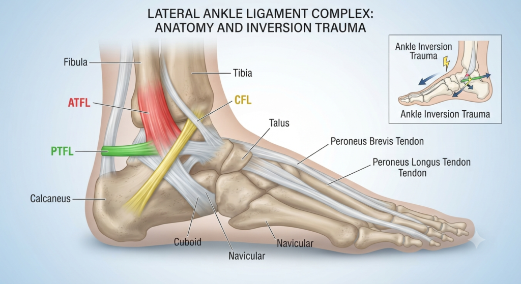
At Al Resalah Medical Center, our multidisciplinary team approaches ankle injuries with a rigorous clinical eye, understanding that inadequate rehabilitation is the leading precursor to Chronic Ankle Instability (CAI), persistent pain, and premature joint degeneration. The lateral ligament complex is most frequently injured, comprising three primary structures:
Anterior Talofibular Ligament (ATFL): The most frequently injured ligament, providing restraint against anterior displacement of the talus and internal rotation when the foot is plantarflexed.
Calcaneofibular Ligament (CFL): Provides stability in both neutral and dorsiflexed positions, resisting inversion tilt of the talus.
Posterior Talofibular Ligament (PTFL): The strongest of the lateral ligaments, rarely injured unless a severe dislocation or high-grade sprain occurs.
2. Classification & Grading of Ankle Sprains
Grade 1 (Mild)
Microscopic stretching of ligament fibers; no macroscopic tear.
Mild localized tenderness, minimal swelling, slight or no functional loss; full weightbearing usually tolerated.
1 to 2 weeks
Grade 2 (Moderate)
Partial macroscopic tear of the ligament (typically ATFL and/or CFL).
Moderate to severe pain, localized swelling, ecchymosis (bruising), moderate joint instability, and antalgic gait.
3 to 6 weeks
Grade 3 (Severe)
Complete rupture of one or more lateral ligaments (ATFL, CFL, and/or PTFL).
Severe immediate pain, extensive swelling, diffuse bruising, profound mechanical instability, and inability to bear weight.
6 to 12+ weeks (Surgical consultation may be indicated)
3. Symptoms and Etiology
Understanding the exact mechanism of injury provides critical diagnostic insight. The overwhelming majority of acute ankle sprains (approximately 85% to 90%) occur via an inversion and plantarflexion mechanism. As the foot rolls underneath the leg, high tensile forces concentrate across the lateral ligament complex.
Common Symptoms
- Acute, sharp pain at the time of injury, transitioning to a dull ache.
- Rapid soft tissue swelling around the lateral malleolus and sinus tarsi.
- Subcutaneous hematoma and ecchymosis tracking down toward the foot.
- Localized tenderness upon palpation of ligamentous insertion sites.
- Mechanical giving way or feeling of joint insecurity.
- Antalgic gait pattern resulting in restricted terminal stance phase.
Primary Etiological Factors
- History of prior ankle sprain (highest predictive risk factor).
- Deficits in proprioceptive feedback and dynamic neuromuscular control.
- Decreased ankle dorsiflexion range of motion (tight gastrocnemius/soleus complex).
- Improper footwear (e.g., narrow heel bases, lack of lateral ankle support during sports).
- Fatigue during high-intensity athletic participation or prolonged standing.
- Structural malalignments such as forefoot varus or hindfoot varus.
4. Clinical Diagnosis & Diagnostic Imaging
A comprehensive physical examination performed by our experienced medical practitioners and neuro-physiotherapists at Al Resalah Medical Center forms the bedrock of accurate diagnosis. Physical assessment includes evaluating active and passive range of motion, neurovascular status, and specific orthopedic stress tests:
- Anterior Drawer Test: Assesses the integrity of the ATFL by translating the calcaneus and talus forward while stabilizing the tibia. Excessive anterior translation compared to the uninjured side indicates a positive test.
- Talar Tilt Test: Evaluates the integrity of the CFL by inverting the calcaneus in a neutral/slightly plantarflexed position.
- Squeeze Test (High Ankle Sprain / Syndesmosis Evaluation): Compresses the tibia and fibula at mid-calf level to rule out syndesmotic (high ankle) injury.
The Ottawa Ankle Rules
To ensure diagnostic efficiency and avoid unnecessary ionizing radiation, our clinicians strictly adhere to the Ottawa Ankle Rules. Radiographs (X-rays) are warranted only if there is pain in the malleolar zone and any of the following: bone tenderness along the distal 6cm of the posterior edge of the tibia/fibula or medial/lateral malleolus, or an inability to bear weight both immediately and for 4 steps in the emergency/clinical setting.
5. Management and Treatment Framework
Modern clinical management has evolved away from strict immobilization toward active, progressive rehabilitation. At Al Resalah Medical Center, our treatment protocol is categorized into three structured phases designed to restore full functional independence.
Phase 1: Acute Protection & Pain Modulation (Days 1 to 7)
The primary objectives in the acute phase are to control hemorrhage, reduce acute neurogenic inflammation, protect healing tissues, and minimize muscle inhibition:
- POLICE Protocol: Protection, Optimal Loading, Ice, Compression, and Elevation. Complete immobilization is avoided unless a Grade 3 instability is confirmed; controlled, pain-free mechanical loading stimulates collagen alignment.
- Modalities: Targeted application of cryotherapy, pulsed shortwave diathermy, or interferential therapy (IFT) to modulate nociceptive input and decrease edema.
- Gentle Mobility: Early introduction of pain-free multi-planar passive range of motion and sub-maximal isometrics.
Phase 2: Restoration of Range of Motion & Neuromuscular Control (Weeks 2 to 4)
Once acute pain subsides, the focus shifts to restoring full joint arthrokinematics and retraining neuromuscular pathways:
- Manual Therapy: Joint mobilizations (posterior-anterior talocrural glides and subtalar distraction) to eliminate joint capsule restrictions and restore normal dorsiflexion.
- Progressive Strengthening: Initiation of elastic resistance bands (TheraBand) across all cardinal planes (plantarflexion, dorsiflexion, inversion, eversion).
- Proprioceptive Training: Single-leg stance drills progressing from firm surfaces to unstable foam pads and wobble boards.
Phase 3: Functional Retraining & Sport-Specific Conditioning (Weeks 5+)
The final phase bridges the gap between clinical rehabilitation and unrestricted functional or athletic performance:
- Plyometric Drills: Linear jumping, multi-directional hopping, and agility ladder drills to restore reactive reflex stabilization.
- Sport-Specific Simulation: Cutting, pivoting, deceleration mechanics, and sport-specific drills under controlled clinical supervision.
- Prophylactic Taping/Bracing: Education on semi-rigid bracing or kinesiology taping for high-risk athletic participation during the initial post-rehab window.
6. Al Resalah Medical Center Physiotherapy Specialists
Our dedicated rehabilitation department features highly qualified clinicians specializing in musculoskeletal, neurological, and sports physiotherapy. Every patient receives an individualized, evidence-based care plan supervised by our senior specialists:


7. Advanced Physiotherapy Treatments
To accelerate cellular healing, reduce recovery times, and ensure superior clinical outcomes, Al Resalah Medical Center utilizes cutting-edge therapeutic technologies:
- Extracorporeal Shockwave Therapy (ESWT): High-energy acoustic waves directed into damaged ligamentous tissue to stimulate neovascularization, accelerate collagen synthesis, and decrease chronic inflammatory markers.
- High-Intensity Laser Therapy (HILT): Penetrates deep into soft tissues to stimulate mitochondrial ATP production, accelerate tissue repair, and provide rapid analgesic relief.
- Computerized Balance & Posturography Testing: Utilizes advanced force-plate technology to quantify center-of-pressure sway, objective postural stability deficits, and asymmetrical weight distribution during rehabilitation.
- Instrument-Assisted Soft Tissue Mobilization (IASTM): Specialized stainless-steel instruments used to break down fascial adhesions, scar tissue, and fibrotic restrictions following trauma.
Clinical Warning: Avoid Premature Return to Activity
A common pitfall in ankle injury recovery is returning to high-impact activities solely because surface pain has subsided. Residual proprioceptive deficits and ligament laxity left unaddressed drastically increase the likelihood of recurrent sprains and early-onset osteoarthritis. Always complete the full functional testing battery under professional guidance.
8. Common Home Exercise Program (HEP)
To support in-clinic rehabilitation, patients are prescribed a customized home exercise program. Consistency is vital for achieving full functional recovery
1. Towel Scrunches (Intrinsic Strengthening)
Execution: Sit upright in a chair with your bare foot resting flat on a small towel placed on a smooth floor. Keeping your heel planted on the ground, use your toes to scrunch the towel toward you. Release and repeat.
Sets/Reps: 3 sets of 10–15 repetitions, twice daily.
2. Alphabet Tracing (Active ROM)
Execution: Sit comfortably with your leg extended or hanging off the edge of a plinth. Imagine your big toe is a pen and trace the letters of the alphabet from A to Z in the air, utilizing your full ankle range of motion.
Sets/Reps: 2 full alphabet sequences, 3 times daily.
3. Resistance Band Eversion (Lateral Stability)
Execution: Sit with legs extended forward. Loop a resistance band around the forefoot of the injured ankle, securing the other end under your healthy foot. Slowly turn your injured foot outward (eversion) against the resistance, then return with control.
Sets/Reps: 3 sets of 12 repetitions.
4. Single-Leg Balance (Proprioception)
Execution: Stand near a supportive wall or countertop for safety. Lift your uninjured foot and balance entirely on the injured ankle. Maintain an upright posture with a micro-knee bend. Progress from eyes open to eyes closed.
Duration: 3 sets of 30 to 60 seconds.
Written by Fayyaz Husain Physiotherapist
Medically Reviewed by Al Resalah Medical Team
Last Reviewed: July 2026