 +971 6 522 6368
  +971-56-994-6523
  info@resalahphysio.com

Comprehensive Guide to Achilles Tendinitis:
Symptoms, Causes, Advanced Diagnosis, and Physical Therapy Interventions
Achilles tendinitis is one of the most common and persistent overuse injuries affecting the lower extremity, particularly among runners, active individuals, and those subject to sudden increases in physical demands. At Al Resalah Medical Center, our multidisciplinary team of rehabilitation specialists, orthopedics, and physiotherapists encounter this condition frequently. Because the Achilles tendon is the largest and strongest tendon in the human body—withstanding loads multiple times your body weight during running and jumping—injuries here require a nuanced, evidence-based approach to recovery
What is Achilles Tendinitis?
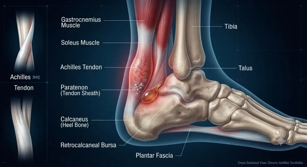
Achilles tendinitis is an inflammatory and degenerative condition affecting the large tendon running down the back of the lower leg, connecting the gastrocnemius and soleus calf muscles to the calcaneus (heel bone). The condition typically manifests in one of two ways based on anatomical location:
Non-insertional Achilles Tendinitis: Characterized by microscopic tears, swelling, and thickening occurring in the middle portion of the tendon fibers. This form is more prevalent in younger, active individuals and athletes.
Insertional Achilles Tendinitis: Involves the lower portion of the tendon where it directly attaches to the heel bone (calcaneus). Bone spurs (osteophytes) frequently develop at this insertion point, and the condition can affect individuals regardless of their activity level.
Primary Causes and Risk Factors
Understanding the mechanical and physiological triggers of Achilles tendinitis is essential for both prevention and long-term rehabilitation. Key factors include:
Sudden Training Spikes: Rapidly increasing the volume, intensity, or frequency of exercise—such as drastically increasing weekly running mileage without adequate adaptation time—overwhelms the tendon’s capacity to remodel.
Calf Muscle Tightness and Weakness: Tight gastrocnemius and soleus complexes place constant passive tension across the Achilles tendon, amplifying the mechanical stress applied during daily ambulation and athletic tasks.
Haglund’s Deformity: An anatomical bony enlargement on the posterior superior aspect of the calcaneus. This bony prominence causes mechanical impingement against the deep bursa and the anterior surface of the Achilles tendon during ankle dorsiflexion, provoking chronic friction and inflammation.
Biomechanical Malalignments: Excessive foot pronation, flat feet (pes planus), or rigid foot structures alter the natural path of load dissipation, increasing torsional forces on the lower leg.
Pharmacological Influences: The systemic use of fluoroquinolone antibiotics is strongly linked with an elevated risk of tendon pathology and spontaneous rupture, demanding extreme caution when prescribing or undertaking exercise regimens while on these medications.
Clinical Presentation and Symptoms
Patients suffering from Achilles tendinitis typically present with a distinct cluster of clinical indicators:
Morning Stiffness and Pain: Sharp pain and marked stiffness along the Achilles tendon upon taking the first steps out of bed, which typically eases after a brief period of gentle movement.
Activity-Related Discomfort: Pain along the tendon or posterior heel that intensifies during physical exertion and plateaus or worsens afterward.
Delayed Post-Exercise Pain: Severe, sharp discomfort presenting 24 hours after a strenuous workout session.
Palpable and Visual Changes: Observable thickening or nodularity within the tendon substance, alongside localized swelling that may persist all day or fluctuate with activity.
Insertional Bone Spurs: Palpable bony prominence at the heel attachment site.
Footwear Intolerance: Localized pain on the back of the heel elicited by the back counter of enclosed shoes or boots.
Advanced Clinical Diagnosis
Accurate diagnosis requires a combination of detailed history-taking, comprehensive physical examination, and, when indicated, diagnostic imaging.
Imaging Modalities
X-Rays: Provide high-resolution images of foot and leg bone structures, identifying calcific tendinopathy, bone spurs, and Haglund’s deformity.
MRI Scans: Excellent for visualizing soft tissue integrity, detecting partial or complete tendon ruptures, and assessing the degree of intratendinous degeneration (tendinosis).
Ultrasound Scans: Dynamic imaging modality that captures real-time tendon movement, structural fiber disruption, hypervascularity, and paratendon inflammation.
Specialized Clinical Tests
To isolate tendon integrity and differentiate specific pathologies, our clinical team relies on targeted physical examinations:
The Thompson Test: Performed with the patient lying prone with feet hanging off the edge of the examination table. The clinician squeezes the calf muscles. A normal, intact tendon causes immediate plantarflexion of the foot. Absence of plantarflexion indicates a complete Achilles tendon rupture.
The Matles Test: The patient lies prone with knees flexed to 90 degrees while keeping the feet relaxed. In a normal, uninjured state, the foot rests in slight plantarflexion due to resting muscle tone. If the foot neutralizes or moves into dorsiflexion, it is a positive test indicating a complete Achilles tendon rupture.
The Royal London Hospital Test: The clinician first palpates the area of maximum tenderness with the patient’s knee extended. The tender area is then re-palpated with the knee flexed to 90 degrees. A marked reduction or total absence of pain in knee flexion is considered a positive sign for Achilles tendinopathy.
Comprehensive Physical Therapy Interventions
Physical therapy forms the foundation of conservative management for Achilles tendinitis, focusing on pain modulation, tissue remodeling, biomechanical correction, and progressive load management.
Acute Management and Modalities
RICE Protocol: Rest, Ice, Compression, and Elevation during acute flare-ups to control localized inflammation and capillary permeability.
Electrotherapy and Adjunct Modalities:
Extracorporeal Shockwave Therapy (ESWT): Utilizes low- or high-energy acoustic shockwaves directed at the damaged tendon tissue to stimulate neovascularization, downregulate chronic inflammation, and accelerate cellular repair. High-energy ESWT is typically completed in a single session (often requiring local anesthesia), whereas low-energy ESWT is spread across 3 to 4 non-anesthetized sessions.
Iontophoresis: Utilizes a mild electrical current to drive anti-inflammatory medications (such as dexamethasone) across the skin barrier, offering modest support primarily in acute presentations.
TENS (Transcutaneous Electrical Nerve Stimulation): Applied to assist with local pain gating.
Rigid Taping and K-Taping: Applied to unload tension off the Achilles tendon and optimize foot posture during functional movement.
Dry Needling: Combined under ultrasound guidance with targeted loading programs to address chronic pain in individuals presenting with symptoms exceeding 3 months and significant tendon thickening.
Acupuncture and Heat Therapy: Used selectively to relax local musculature and ease myofascial restriction.
Night Splinting: Removable nocturnal braces hold the ankle in a neutral or slightly dorsiflexed position throughout the night. This maintains calf muscle flexibility and prevents the acute morning shortening and subsequent tensile strain when first stepping out of bed.
Structured Stretching and Flexibility Protocols
To restore normal physiological compliance to the gastrocnemius-soleus-Achilles complex, clinicians prescribe structured, progressive stretches:
+---------------------------------------------------------------------------------+
|                         ESSENTIAL STRETCHING ROUTINE                            |
+--------------------------+------------------------------------------------------+
| Stretch Name             | Protocol Summary                                     |
+--------------------------+------------------------------------------------------+
| Towel Stretch            | Sit with leg extended; loop towel around ball of foot|
|                          | and pull toward body; hold 30s, repeat 3x.           |
+--------------------------+------------------------------------------------------+
| Gastrocnemius Stretch    | Staggered stance against wall, back knee straight,   |
|                          | heel down; hold 30s, repeat 3-5x daily.              |
+--------------------------+------------------------------------------------------+
| Soleus Stretch           | Staggered stance, back knee slightly bent, shifting  |
|                          | weight downward into lower calf; hold 30s.           |
+--------------------------+------------------------------------------------------+
| Plantar Fascia Stretch   | Ball of foot on stair, dropping heel below step      |
|                          | level; hold 30s, repeat 3x.                          |
+--------------------------+------------------------------------------------------+
| Toe Stretch              | Seated, extend leg with heel on floor, pull big toe  |
|                          | up and back toward ankle; hold 15-30s.               |
+--------------------------+------------------------------------------------------+
6. Al Resalah Medical Center Physiotherapy Specialists
Our dedicated rehabilitation department features highly qualified clinicians specializing in musculoskeletal, neurological, and sports physiotherapy. Every patient receives an individualized, evidence-based care plan supervised by our senior specialists:


Progressive Strengthening and Neuromuscular Control Exercises
Rehabilitation must progress from isometric loading to isotonic concentric and eccentric loading to rebuild structural tensile strength:
Toe Raises: Standing in a normal weight-bearing posture, rock backward onto your heels so that the forefeet and toes elevate off the floor. Hold for 5 seconds; complete 3 sets of 10 repetitions.
Heel Raises: Standing behind a stable support, rise upward onto your toes, hold for 5 seconds, and slowly lower back down. Complete 3 sets of 10 repetitions.
Single-Leg Balance: Stand unassisted on the injured limb with eyes open, progressing to eyes-closed conditions to challenge neuromuscular stability. Hold for 30 seconds; repeat 3 times.
Side-Lying Leg Lift: Lying on the uninjured side with the front thigh muscles engaged, lift the injured leg 8 to 10 inches upward while maintaining a straight knee. Complete 2 sets of 15 repetitions.
Step-Ups: Place the injured foot on a 3- to 5-inch platform. Shift your body weight onto the support leg, straightening it as the uninjured leg lifts off the floor, then slowly lower back down. Complete 2 sets of 15 repetitions.
Balance and Reach Exercises: Standing on the injured leg next to a chair with a slightly flexed knee, reach forward or across the body to challenge dynamic core stability and lower-extremity control. Perform 2 sets of 15 repetitions.
Interventional Procedures and Medical Management
When conservative physical therapy fails to resolve chronic, recalcitrant tendinopathy, medical interventions may be considered:
Sclerosing Injections: Targeted treatments designed to address abnormal neovascularization and accompanying nerve ingrowth associated with chronic pain.
Corticosteroid Injections: Deployed selectively under strict medical supervision (keeping in mind the inherent risks of structural weakening within weight-bearing tendons).
Non-Steroidal Anti-Inflammatory Drugs (NSAIDs): Used judiciously for short-term pain relief during acute inflammatory phases.
Preventive Guidelines and Lifestyle Modification
Long-term management of Achilles tendinitis requires sustained adherence to preventive mechanics:
Footwear Selection: Choose shoes featuring robust cushioning, optimal arch support, and a slightly elevated heel drop to reduce immediate tension on the Achilles complex.
Gradual Heel Transition: When transitioning from high-heeled footwear to flat shoes, reduce heel height gradually to allow the tendon to adapt its resting length.
Surface Modification: Perform running and athletic activities on flat, even surfaces whenever possible.
Medication Awareness: Exercise caution and consult your physician if engaging in high-impact training while taking fluoroquinolone class antibiotics.
Active Rest: Immediately halt activities that reproduce sharp tendon pain, allowing adequate biological tissue recovery.
At Al Resalah Medical Center, our dedicated team is committed to providing comprehensive, patient-centered care for all musculoskeletal conditions. If you are experiencing persistent heel or lower leg discomfort, contact our clinic today to schedule a comprehensive evaluation with our specialized physiotherapy team.
Written by Fayyaz Husain Physiotherapist
Medically Reviewed by Al Resalah Medical Team
Last Reviewed: July 2026